Hear the Code of Life
Icosmo Helix translates DNA, RNA and the proteins they code for into sound and colour through the Icosmo Matrix. Every building block of life vibrates at its own measured molecular frequency: the four bases, their carbonyl groups, the hydrogen bonds between the two strands, the phosphates, links and sugars of the backbone – and now also the 20 amino acids and the peptide bond that joins them into a protein. Helix brings these vibrations into the audible range by octaves and plays any gene as it is written: base by base, codon by codon, strand by strand, amino acid by amino acid.
Choose a gene from the library – human insulin, the language gene FOXP2, the pigment gene of a lion, the photosynthesis of a plant – search for any animal or plant by its name, or enter your own sequence. Watch the double helix turn with the music, hear the mRNA being translated into protein, see the tones as patterns in a virtual water bowl, follow every codon and its amino acid in the sequencer and the genetic code, and mix the layers of the molecule like the channels of a studio. Take the gene with you as MP3, video reel or PDF report.
How to use:
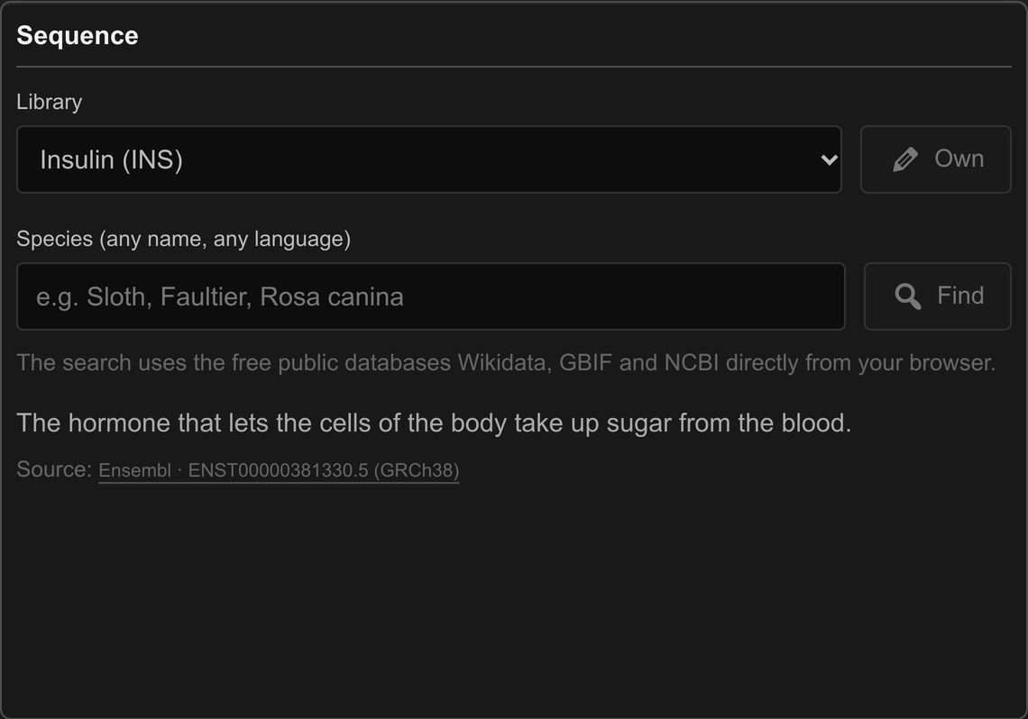
- Choose a gene from the Library, or click Own to paste your own DNA (A, C, G, T) or RNA (A, C, G, U) sequence, also in FASTA format. Ambiguous letters such as N, R or Y are kept in place as N: they stay silent, and the reading frame of the gene is not disturbed. Your sequence stays in your browser.
- Or type any animal or plant into Species – in any language, typos allowed – and press Find. Helix loads its DNA barcode gene.
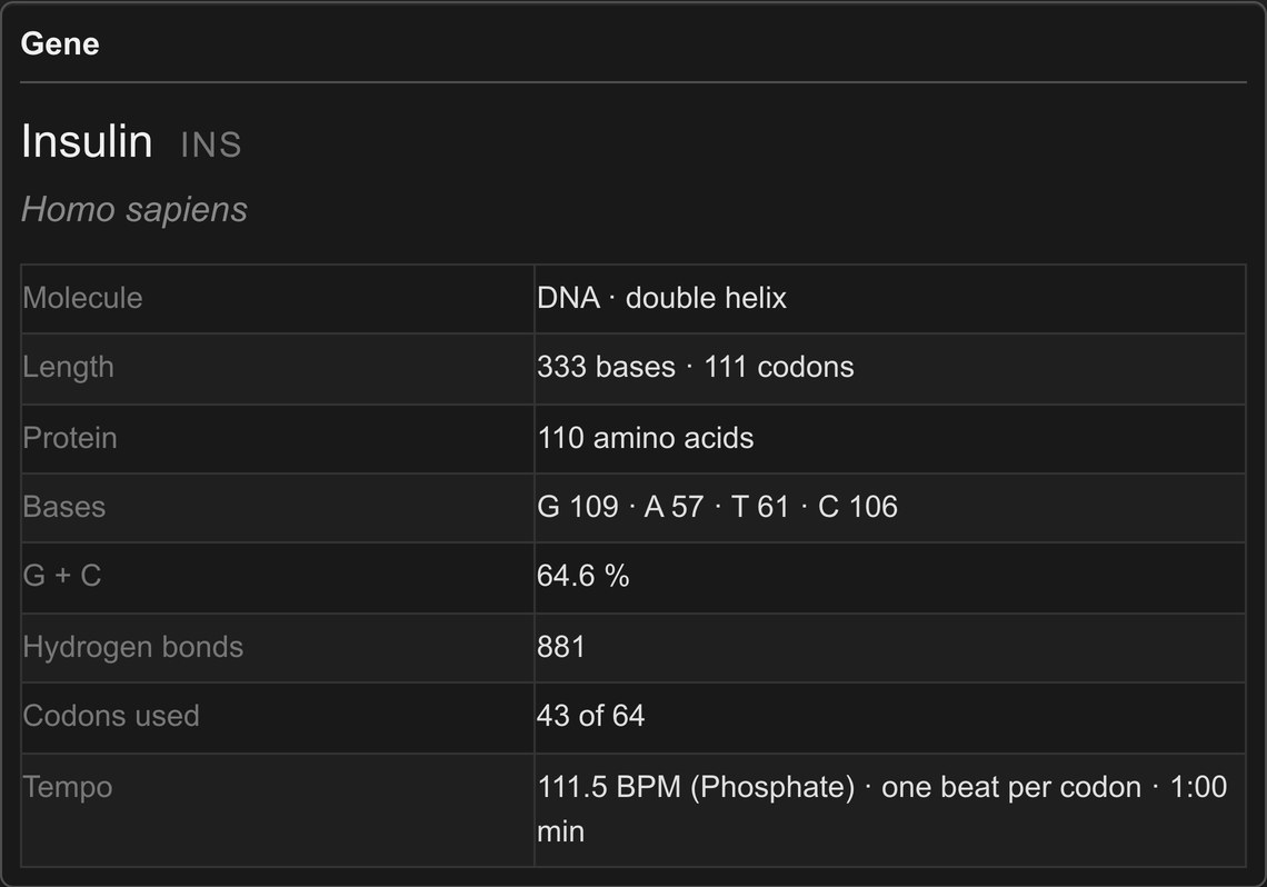
- The Gene panel shows what you are listening to: length, protein, bases, G + C content, hydrogen bonds, the genetic code used and the duration at the tempo of the phosphate.
- Press Play to hear the gene from the selected codon. Press it again to stop. The Loop button plays the gene again and again.
- Switch the Molecule between DNA, the gene as it is stored, and mRNA, the working copy the cell translates into protein.
- Choose the beat: One beat = Codon plays the three bases of each codon as a triplet; One beat = Nucleotide gives every base the full beat.
- Choose the sound: the sine symbol lets the structure of the molecule sound as standing waves, the drum symbol turns it into rhythm.
- Mix the layers with the faders. In the Sequencer, click the upper half of a step to jump to a codon, or the lower half to hear its amino acid. Click a row of the legend or a card at the bottom of the page to hear a single building block.
- Save the gene with Export as MP3, Video Reel or PDF Report.
Note: All calculations, all sound and all exports are made locally in your browser. No sequence is sent to a server. The species search asks the free public databases Wikidata, GBIF and NCBI directly from your browser.
Decoding the Helix
Every part of the Helix player shows one layer of the molecule and its place in the Icosmo Matrix. Here is what each part does:
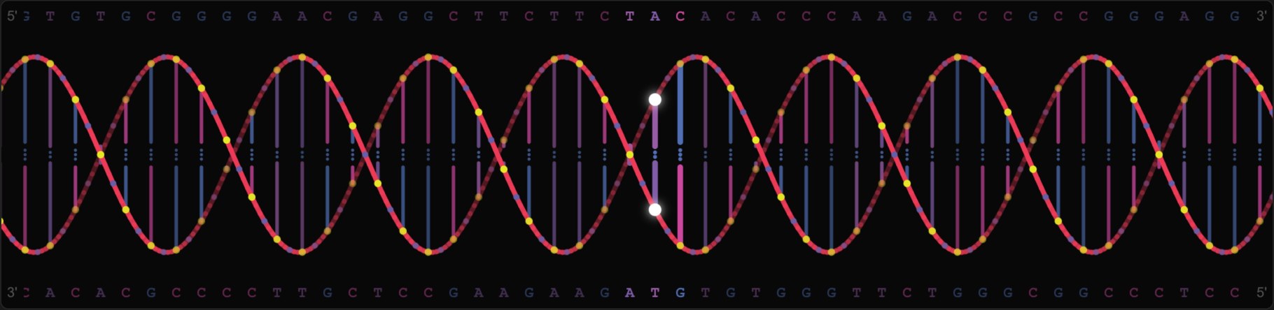
The Double Helix:
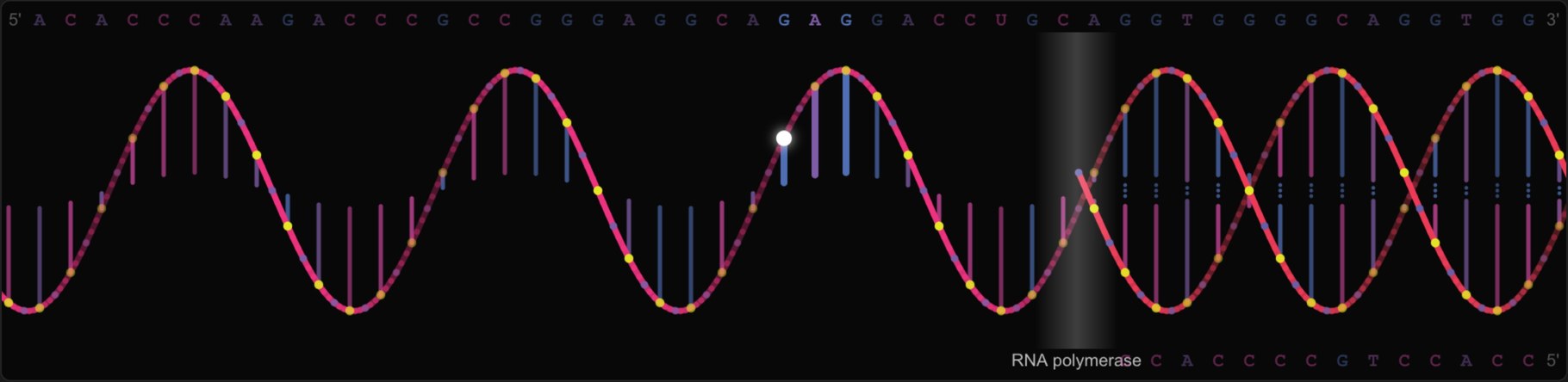
DNA is drawn as the ladder it is. Each rung is a base pair in the colours of its two bases, with the hydrogen bonds that hold it together shown as two dots (Adenine–Thymine) or three dots (Guanine–Cytosine). The rails are the backbone: the phosphodiester link in its own colour, a yellow dot for every phosphate and a blue-violet dot for every sugar between them, exactly as sugar and phosphate alternate in the real molecule. The coding strand runs from 5′ to 3′ along the top, its partner strand along the bottom. The helix only moves while the gene is playing and follows the codon you hear.
Water and Oscilloscope:
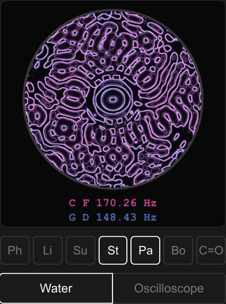
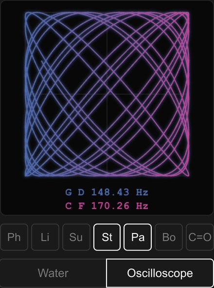
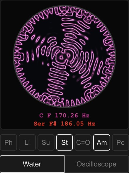
Next to the helix, the tones of the moment become visible. Choose which building blocks take part with the buttons below the image. In DNA these are Ph (phosphate), Li (link), Su (sugar), St (strand), Pa (pair), Bo (hydrogen bond) and C=O (carbonyl). In mRNA, Pair and Bond give way to Am (the amino acid of the codon) and Pe (the peptide bond). Switch between two views:
- Water – cymatics in a virtual water bowl, 4 cm wide and 1 cm deep. The selected tones shake the bowl like a loudspeaker beneath it, and the water answers at half their frequency, as Michael Faraday first described in 1831 (Faraday waves). The physics of water waves gives the wavelength of every tone; waves run in from the wall, are reflected, cross each other, and standing patterns grow. A ring lamp above draws a thin line where the water tilts, like the water sound photographs of Alexander Lauterwasser. Every tone has its own pattern, and several tones together weave an interference pattern that keeps changing while the gene plays. The faders set how strongly each tone shakes the water.
- Oscilloscope – the strand moves a point from left to right, all other selected tones together from top to bottom. The ratios of the frequencies draw the figure: simple ratios close into calm loops, complex ones weave dense patterns.
The water bowl is a simplified physical model. Real experiments also depend on the volume, the material of the bowl and nonlinear effects.
From DNA to mRNA to Protein:
Every living being – human, animal, plant, fungus or bacterium – stores its genes as DNA and uses them by making copies as RNA. Switch to mRNA and Helix shows the messenger RNA of the same gene: a single strand without a partner, with Uracil (U) in place of Thymine, its own sugar (ribose) and its own backbone link. You can see the change as a transcription: a glowing reading front, the RNA polymerase, runs along the gene, opens the double strand and leaves the single mRNA strand behind it.
And you can hear it: the pair strand and the hydrogen bonds fall silent, the backbone moves from the key of F# to the key of F – and the translation begins. As the ribosome does in the cell, Helix reads the mRNA codon by codon: the Amino layer plays the amino acid of every codon at its own frequency, and the Peptide layer strikes once for every peptide bond that joins a new amino acid to the growing protein.
The Mixer:
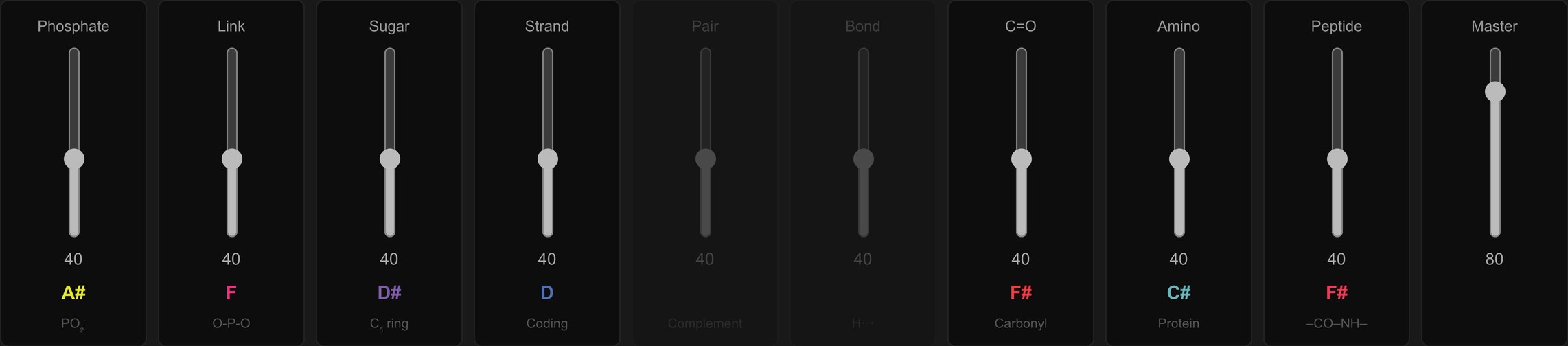
Each fader controls one building block of the molecule and shows its key in its Matrix colour:
- Phosphate (A#) – the beat of the backbone. Its tempo of 111.5 BPM is the clock of the whole player.
- Link (F# in DNA, F in RNA) – the phosphodiester bond between two nucleotides.
- Sugar (D#) – deoxyribose in DNA, ribose in RNA.
- Strand – the bases of the gene, each at its own frequency.
- Pair – the complementary bases on the second strand (DNA only).
- Bond (D) – the hydrogen bonds between the pairs (DNA only).
- C=O (F#) – the carbonyl groups of Thymine and Uracil.
- Amino – the amino acid of every codon, held for the whole codon (mRNA only). Its key changes with every amino acid.
- Peptide (F#) – the peptide bond that joins the amino acids into a protein (mRNA only).

Rhythm or Standing Wave:
With the drum symbol, the structure of the molecule becomes rhythm: the phosphate pulses on every beat, the link and the sugar pulse in turn for every nucleotide, the hydrogen bonds strike two or three times for every pair, the carbonyl groups sound with their base, and in mRNA the peptide bond strikes for every new amino acid. Fade out Strand and Pair, and you hear the pure beat pattern of the gene.
With the sine symbol, every building block of the structure holds its frequency as a standing wave, and only the bases and amino acids move. Together they form the chord of the molecule – all participating frequencies at once.
The Sequencer and the Genetic Code:
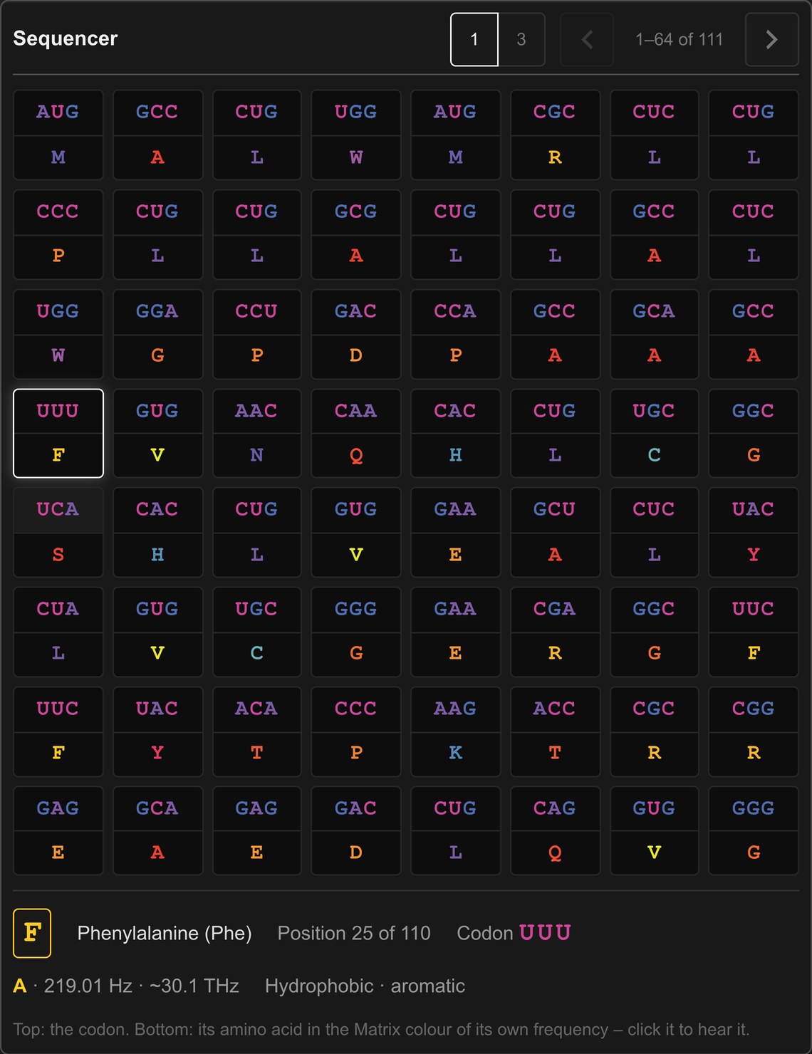
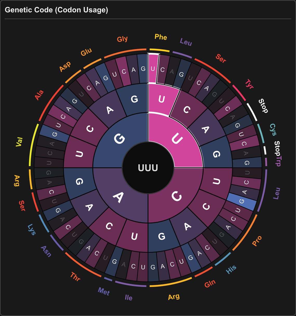
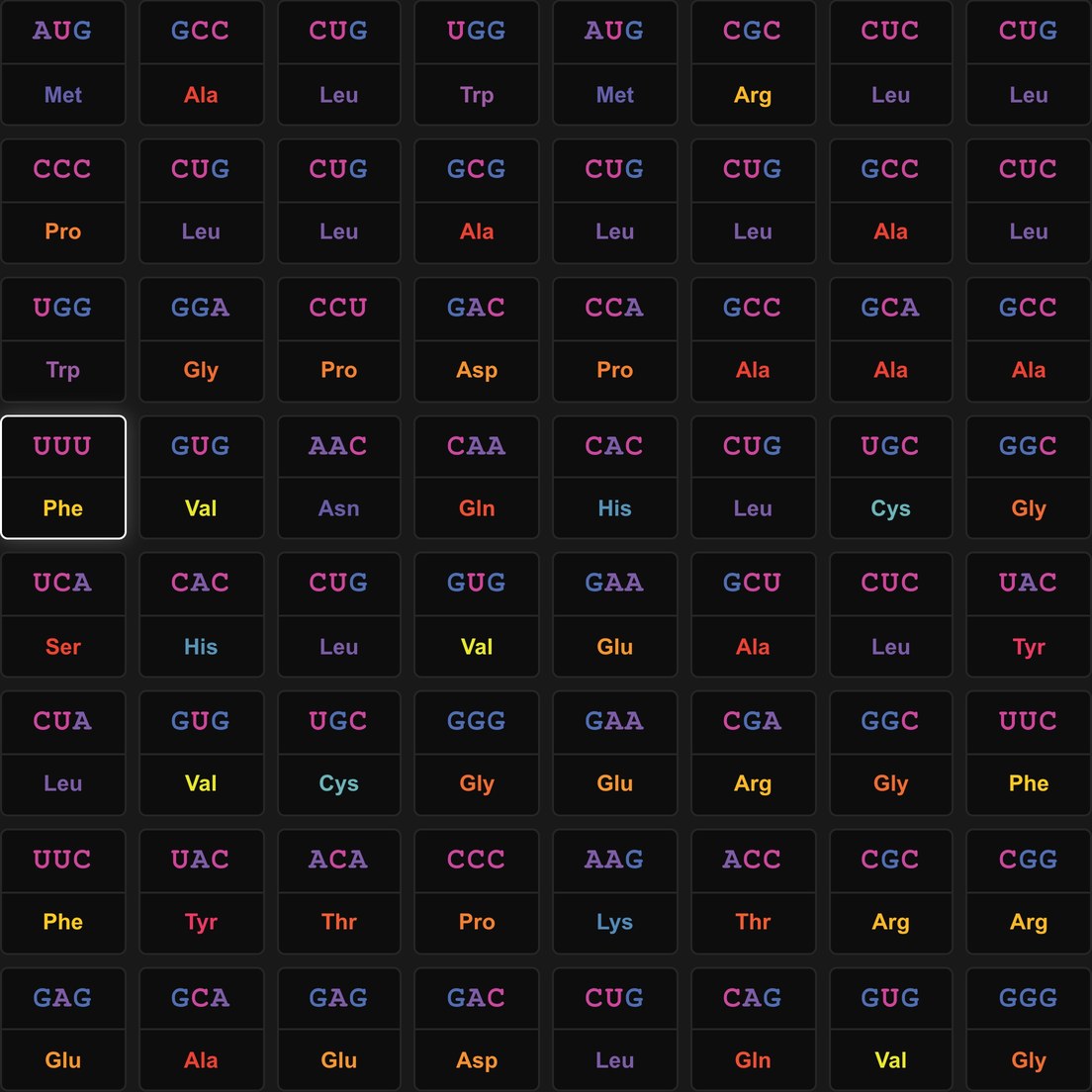
The genetic code is read in words of three letters, the codons. There are 4 × 4 × 4 = 64 of them, and each one stands for one of 20 amino acids or for the stop signal. The Sequencer shows the gene in pages of 64 steps. Each step has two halves: the codon on top, in the colours of its bases, and its amino acid below, in the Matrix colour of the amino acid’s own frequency. Switch between one-letter codes (1, e.g. F) and three-letter codes (3, e.g. Phe). Below the steps, the selected amino acid is described: name, position in the protein, codon, key, tone and chemical character.
The Genetic Code disk shows all 64 codons in three rings – first, second and third base – with the amino acids around the outside in their Matrix colours. It glows where the gene uses a codon most often and marks the codon being played. The stop signal is not a molecule and has no frequency, so it is shown in neutral white.
Mitochondria and chloroplasts read some codons differently; Helix uses the genetic code of the organism, as published by NCBI.
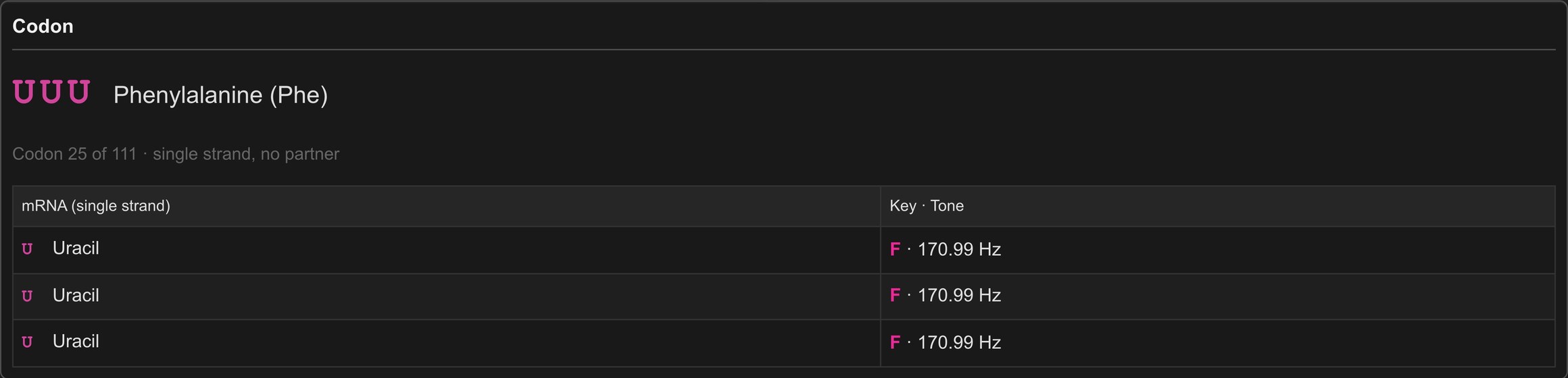
The Codon:
The Codon panel shows every base of the codon being played, its partner on the second strand (in DNA), the number of hydrogen bonds, and the key and exact tone of each base.
Any Animal, Any Plant:
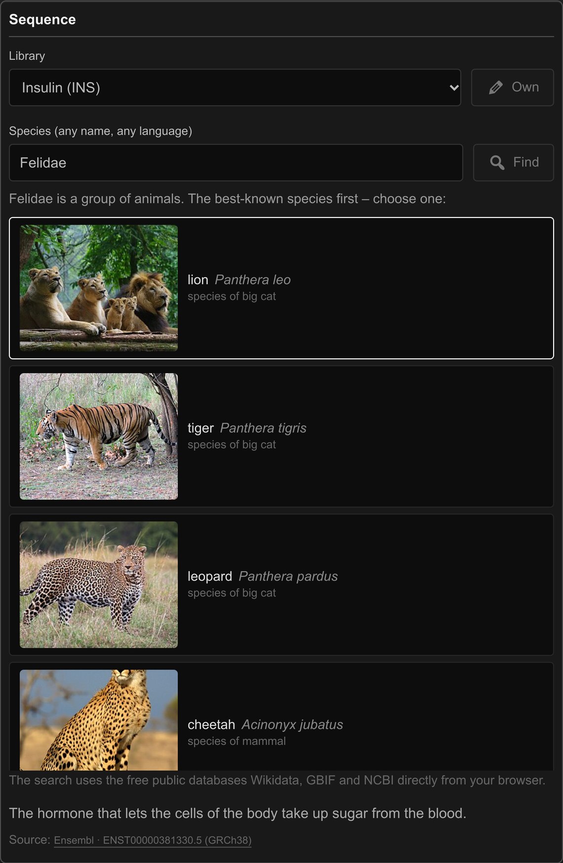
Type the name of any animal or plant into Species – sloth, Faultier, perezoso or Folivora, even with a typo. Helix finds it in Wikidata, checks in the NCBI Taxonomy and in GBIF whether it is an animal or a plant, and loads its DNA barcode from NCBI GenBank: the gene science uses to tell species apart. For animals this is COI from the mitochondrion, the power plant of the cell; for plants it is rbcL from the chloroplast, the gene of photosynthesis.
Mitochondria and chloroplasts read their genes with their own variants of the genetic code, and Helix uses the right one for every organism. If you search a group – roses, cats, whales – Helix shows its best-known species first, ranked by how many Wikipedia articles exist about them, each with its photo from Wikimedia Commons.

Export:
Take the gene with you. Everything is rendered on your own device and nothing is uploaded:
- MP3 – the gene from the selected codon, up to 5 minutes, with your mix, beat and sound.
- Video Reel – 60 seconds in 9:16 for Instagram, TikTok and YouTube Shorts: the turning helix, the water bowl or oscilloscope, the photo of the species and every codon, with its sound. It needs a current browser that can encode video.
- PDF Report – the gene on paper: its data, the photo, the genetic code disk and the Matrix tones of all building blocks.
An export that is running can be stopped at any time with Cancel.
The Library
All genes in the library are real coding sequences from the free public databases Ensembl, Ensembl Plants and NCBI GenBank, each with a link to its source:
- Human – insulin, haemoglobin, oxytocin, the pigment gene MC1R and the language gene FOXP2.
- Human Populations – the skin pigment gene SLC24A5 of ten real people from the 1000 Genomes Project: a woman and a man from Africa, Europe, East Asia, South Asia and the Americas. All ten share this gene letter for letter – except for a single base at codon 111, the one letter most strongly linked to the colour of human skin.
- Animal and Plant – the lion, the glowing jellyfish, the chimpanzee and the photosynthesis genes of a flowering plant.
- Insulin across Species and MC1R across Species – the same gene in human, mouse, cow, pig, chicken, zebrafish, lion, cat and horse. Listen to how one melody changes along the tree of life.
Beyond the library, the Species search opens the DNA barcodes of hundreds of thousands of animals and plants.
Measured, Not Invented
The frequencies of Icosmo Helix are not chosen for musical effect. Each one is a molecular vibration measured by Raman spectroscopy, brought into the audible range by octaves exactly as the Icosmo Calculator does it, and placed in the Icosmo Matrix with its key, colour, tempo and chakra. The four bases of DNA fall on four neighbouring keys – D, D#, E and F – and form one continuous band of colour from blue to red-violet.
The 20 amino acids follow one strict rule: each is tuned by the strongest Raman band of the solid amino acid between 450 and 1800 cm⁻¹, the band spectroscopists use to recognise it. The peptide bond is tuned by its amide I band (1668 cm⁻¹, the median of 68 protein spectra). One result stands out: the peptide bond sounds exactly one octave above the phosphodiester link of DNA. The link that holds a protein together and the link that holds the gene that codes for it share one tone, F#.
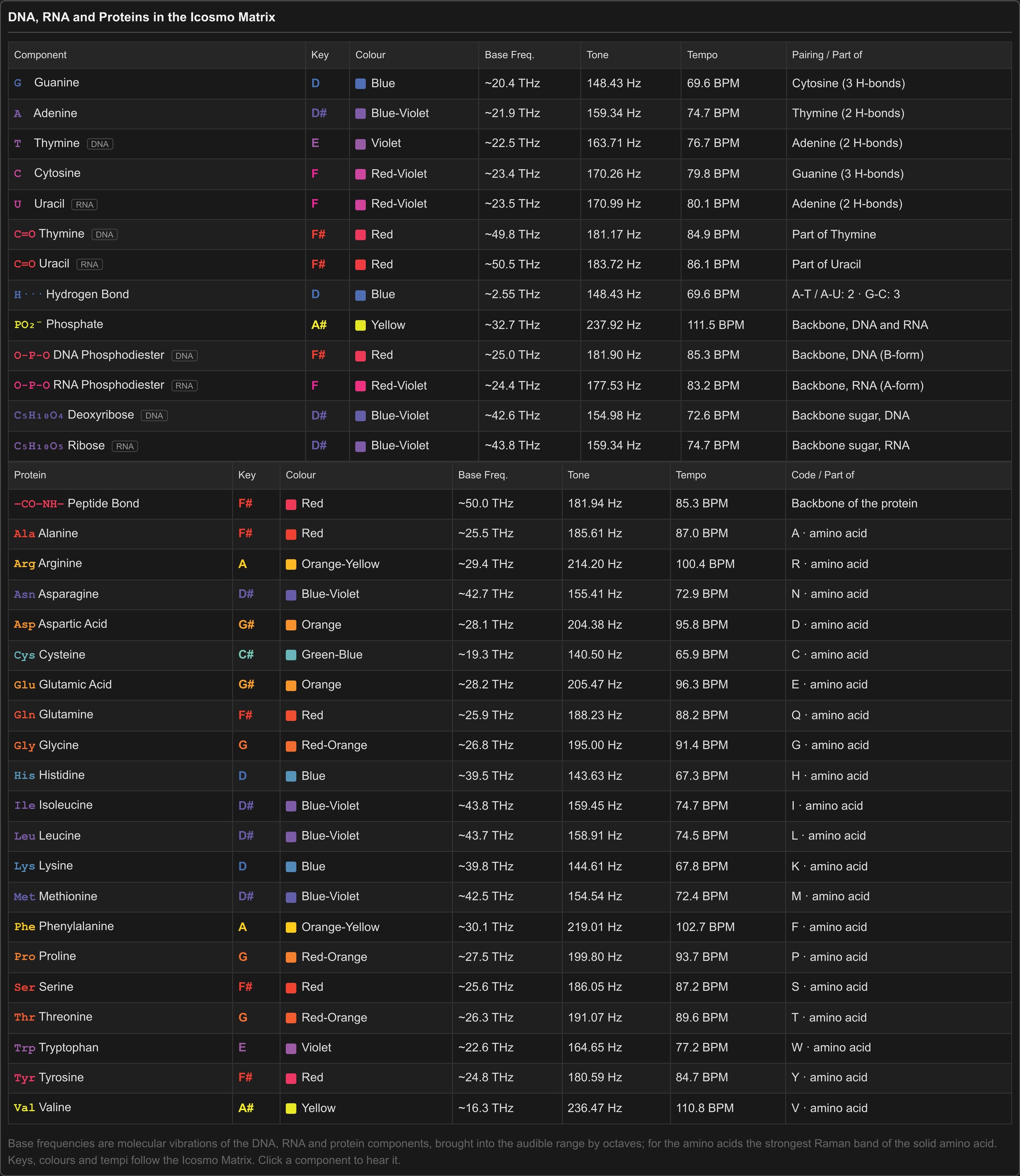
Every building block of DNA and RNA has its own card in the Icosmo DNA Tarot, where you can read its meaning and see its full Matrix data. Cards for the amino acids and the peptide bond will follow.
Sources and References
Icosmo Helix is built on published measurements and free public data. These are the sources of its frequencies, its genetic code and its sequences.
Molecular vibrations of DNA and RNA
- W. Safar, A. Azziz, M. Edely, M. Lamy de la Chapelle: Conventional Raman, SERS and TERS Studies of DNA Compounds. Chemosensors 11 (2023) 399. doi:10.3390/chemosensors11070399 – review of the Raman bands of the DNA bases, the B-DNA backbone link and the phosphate.
- J. M. Benevides, A. H. Wang, G. A. van der Marel, J. H. van Boom, A. Rich, G. J. Thomas Jr.: The Raman spectra of left-handed DNA oligomers incorporating adenine–thymine base pairs. Nucleic Acids Research 12 (1984) 5913–5925. doi:10.1093/nar/12.14.5913 – marker bands of the bases in DNA.
- G. J. Thomas Jr., J. M. Benevides, S. A. Overman, T. Ueda, K. Ushizawa, M. Saitoh, M. Tsuboi: Polarized Raman spectra of oriented fibers of A DNA and B DNA. Biophysical Journal 68 (1995) 1073. doi:10.1016/S0006-3495(95)80282-1 – backbone bands of the A-form (RNA) and the B-form (DNA).
- A. Avni, A. Joshi, A. Walimbe, S. G. Pattanashetty, S. Mukhopadhyay: Single-droplet surface-enhanced Raman scattering decodes the molecular determinants of liquid-liquid phase separation. Nature Communications 13 (2022) 4378. doi:10.1038/s41467-022-32143-0 – the ring breathing vibration of Uracil in RNA.
- H. Urabe, Y. Tominaga: Low-lying collective modes of DNA double helix by Raman spectroscopy. Biopolymers 21 (1982) 2477–2481. doi:10.1002/bip.360211212, and H. Urabe, Y. Tominaga, K. Kubota: Experimental evidence of collective vibrations in DNA double helix. The Journal of Chemical Physics 78 (1983) 5937–5939. doi:10.1063/1.444600 – the low-frequency vibration of the hydrogen bonds between the base pairs.
Molecular vibrations of the amino acids and the peptide bond
- J. De Gelder, K. De Gussem, P. Vandenabeele, L. Moens: Reference database of Raman spectra of biological molecules. Journal of Raman Spectroscopy 38 (2007) 1133–1147. doi:10.1002/jrs.1734 – Glycine, Alanine, Arginine, Glutamic acid, Histidine, Phenylalanine, Proline, Serine, Tryptophan, Tyrosine and Valine. Read from the open peak database RamanBiolib.
- J. Casado, J. T. López Navarrete, F. J. Ramírez: Infrared and Raman spectra of L-asparagine and L-asparagine-d5 in the solid state. Journal of Raman Spectroscopy 26 (1995) 1003–1008. doi:10.1002/jrs.1250261105 – Asparagine.
- J. T. López Navarrete, V. Hernández, F. J. Ramírez: IR and Raman spectra of L-aspartic acid and isotopic derivatives. Biopolymers 34 (1994) 1065–1077. doi:10.1002/bip.360340810 – Aspartic acid.
- SDBS: https://sdbs.db.aist.go.jp/, National Institute of Advanced Industrial Science and Technology (AIST), Japan, accessed 15 October 2026 – Glutamine, Isoleucine and Leucine (Raman spectra by K. Tanabe and J. Hiraishi).
- M. Zhezhu, A. Vasil’ev, H. Parsamyan, T. Abrahamyan, G. Baghdasaryan, S. Gyozalyan, D. A. Ghazaryan, H. Gharagulyan: L-Cysteine Polymorph Coatings for THz Sensing Metasurfaces. arXiv (2025). arXiv:2507.20209 – Cysteine.
- J. G. de Oliveira Neto, O. C. da Silva Neto, J. A. O. Rodrigues, J. R. Viana, A. Steimacher, F. Pedrochi, F. F. de Sousa, A. O. dos Santos: Effect of Dy³⁺ Ions on Structural, Thermal and Spectroscopic Properties of L-Threonine Crystals. Quantum Beam Science 9 (2025) 3. doi:10.3390/qubs9010003 – Threonine.
- V. S. Gorelik, V. P. Sverbil, B. P. Gorshunov, A. I. Seredin: Pseudoscalar lattice modes in the amino acid crystals and DNA. Journal of Physics: Conference Series 918 (2017) 012033. doi:10.1088/1742-6596/918/1/012033 – Lysine.
- P. T. C. Freire, F. M. Barboza, J. A. Lima, F. E. A. Melo, J. Mendes Filho: Raman Spectroscopy of Amino Acid Crystals. In: Raman Spectroscopy and Applications, IntechOpen (2017). doi:10.5772/65480 – Methionine.
- A. Rygula, K. Majzner, K. M. Marzec, A. Kaczor, M. Pilarczyk, M. Baranska: Raman spectroscopy of proteins: a review. Journal of Raman Spectroscopy 44 (2013) 1061–1076. doi:10.1002/jrs.4335 – most of the protein spectra from which the amide I band of the peptide bond was taken (via RamanBiolib).
Genetic code, sequences and species
- The Genetic Codes (NCBI translation tables) – the standard code and the codes of mitochondria and plastids, with their start codons.
- Ensembl and Ensembl Plants – the coding sequences of the library.
- 1000 Genomes Project – the SLC24A5 sequences of the Human Populations.
- NCBI GenBank and the NCBI Taxonomy – the DNA barcode genes and the genetic code of every species.
- Wikidata, GBIF and Wikimedia Commons – the names of species in all languages, their place in the tree of life and their photos.
Water and sound
- M. Faraday: On a peculiar class of acoustical figures; and on certain forms assumed by groups of particles upon vibrating elastic surfaces. Philosophical Transactions of the Royal Society of London 121 (1831) 299–340. doi:10.1098/rstl.1831.0018 – the waves that answer at half the driving frequency.
- The water sound photographs of Alexander Lauterwasser – the model for the ring lamp of the water bowl.
- Open-source software running in your browser: Tone.js (sound), jsPDF (PDF), mp4-muxer (video) and lamejs (MP3).
The full research behind the amino acid frequencies – the rule, every value and its source – is part of the Icosmo Matrix research.